This technical report introduces a method for measuring the three-dimensional articular motions of the shoulder complex during swimming. Eleven collegiate swimmers performed front-crawl strokes at maximal effort and their shoulder motions were measured with an electromagnetic tracking device. Sensors were attached to the sternum, acromia and humeri to determine their relative positions and orientations. A cart carrying the components of the device was pushed back-and-forth along the poolside, so that the sensors attached to the swimmer could be detected within the electromagnetic field. The stroke-to-stroke reliability of the measured articular motions was determined for each swimmer. The accuracy of the device was tested by measuring the relative positions and orientations of multiple sensors fixed on a wooden stick moving above and below the water surface. The measured values were compared with pre-determined fixed values. The coefficient of variance for the joint angles between stroke cycles was <10% of the total range of movement. Within a range of 1282 mm from the transmitter, the root-mean-square error of measurement was 0.7° for orientation and 4 mm for position, both of which were superior to optical measurements. This method is accurate and reliable for measuring the kinematics of the shoulder complex during swimming.

KEYWORDS: Electromagnetic tracking device; methodology; kinematics


Purpose: This study aimed to elucidate growth pattern of mechanical properties of the Achilles tendon and to examine if imbalance between growth of bone and muscle–tendon unit occurs during adolescence.

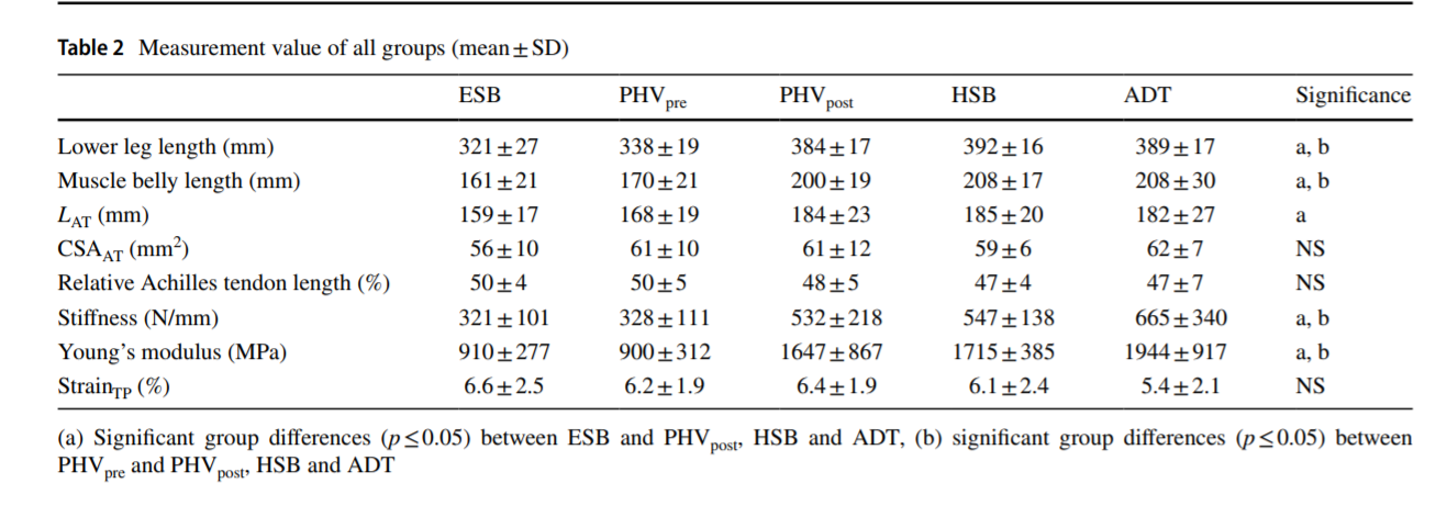
Methods: Fourteen elementary school boys, 30 junior high school boys, 20 high school boys and 15 male adults participated in this study. Based on estimated age at peak height velocity (PHV), junior high school boys were separated into two groups (before or after PHV). An ultrasonography technique was used to determine the length, cross-sectional area, stiffness and Young’s modulus of Achilles tendon. In addition, the maximum strain in “toe region” (strainTP) was determined to describe the balance between growth of bone and muscle–tendon unit.

Results: No group difference was observed in length, cross-sectional area and strainTP among the groups. However, stiffness and Young’s modulus in after PHV groups were significantly higher than those of elementary school boys and before PHV groups (p ≤ 0.05).

Conclusions: These results indicate that mechanical properties of Achilles tendon change dramatically at and/or around PHV to increased stiffness. The widely believed assumption that muscle–tendon unit is passively stretched due to rapid bone growth in adolescence is not supported.

KEYWORDS: Growth, Stiffness, Young’s modulus, Muscle–tendon imbalance, Peak height velocity


本研究では,肩を構成する関節(肩甲上腕関節と肩甲胸郭関節)に着目して,上腕到達範囲において若年者と高齢者との間に生じる差の要因を調べました. 日常的な肩の痛みが無く,20・30代の男性である若年者群51名および60・70代の男性である高齢者群27名が実験に参加しました. 利き腕側の肩を対象とし,可能な限り大きなCircumductionを行わせた際に上腕が到達する範囲を測りました. このとき上腕骨長を一定とし,肩関節中心を基準に上腕骨遠位端が描いた軌跡に囲まれた領域の面積を上腕到達範囲, 肩甲骨を基準に観測したものを肩甲上腕関節の上腕到達範囲への貢献とし,上腕到達範囲と肩甲上腕関節の貢献との差分を肩甲胸郭関節の上腕到達範囲への貢献として求めました. 上腕到達範囲を前額面・水平面によって前後・上下の4領域に分け,高齢者群と若年者群を比べると,高齢者群の若年者群に対する上腕到達半脾の狭まりが上後方の領域において顕著であることがわかりました. また,その領域の差の95%が肩甲骨の貢献によるものでした. したがって,胸郭上後方への到達範囲における若年者群と高齢者群との差の要因は,上腕到達範囲に対する肩甲骨の貢献の低下であることが明らかとなりました.